Lower back pain is a common issue that affects many of us worldwide, often caused by poor posture, muscle imbalances, sedentary lifestyles, injuries, or chronic conditions. While rest and medication may provide temporary relief, long-term recovery requires active rehabilitation—and this is where a Biokineticist plays a crucial role.
How Biokineticists Tackle Lower Back Pain
Assessment & Diagnosis
A Biokineticist first evaluates the root cause of your pain by analysing:
Posture & movement patterns (e.g., sitting posture, Functional movement analysis)
Muscle imbalances (weak core, tight hip flexors, weak glutes)
Mobility & stability
Previous injuries or medical conditions
The above can help in identifying what could be the underlying cause of someone’s back pain, and they are crucial in planning a rehabilitation programme. Not everyone is the same, and individualisation ensures that the person gets the best possible results.
Daily habits (avoiding prolonged sitting, incorporating movement breaks).
Unlike passive treatments that only mask pain, Biokineticists provide long-term solutions by addressing the underlying causes of lower back pain through movement correction, strength training, and education. If you suffer from persistent back pain, consulting a Biokineticist could be the key to lasting relief and a stronger, pain-free back.
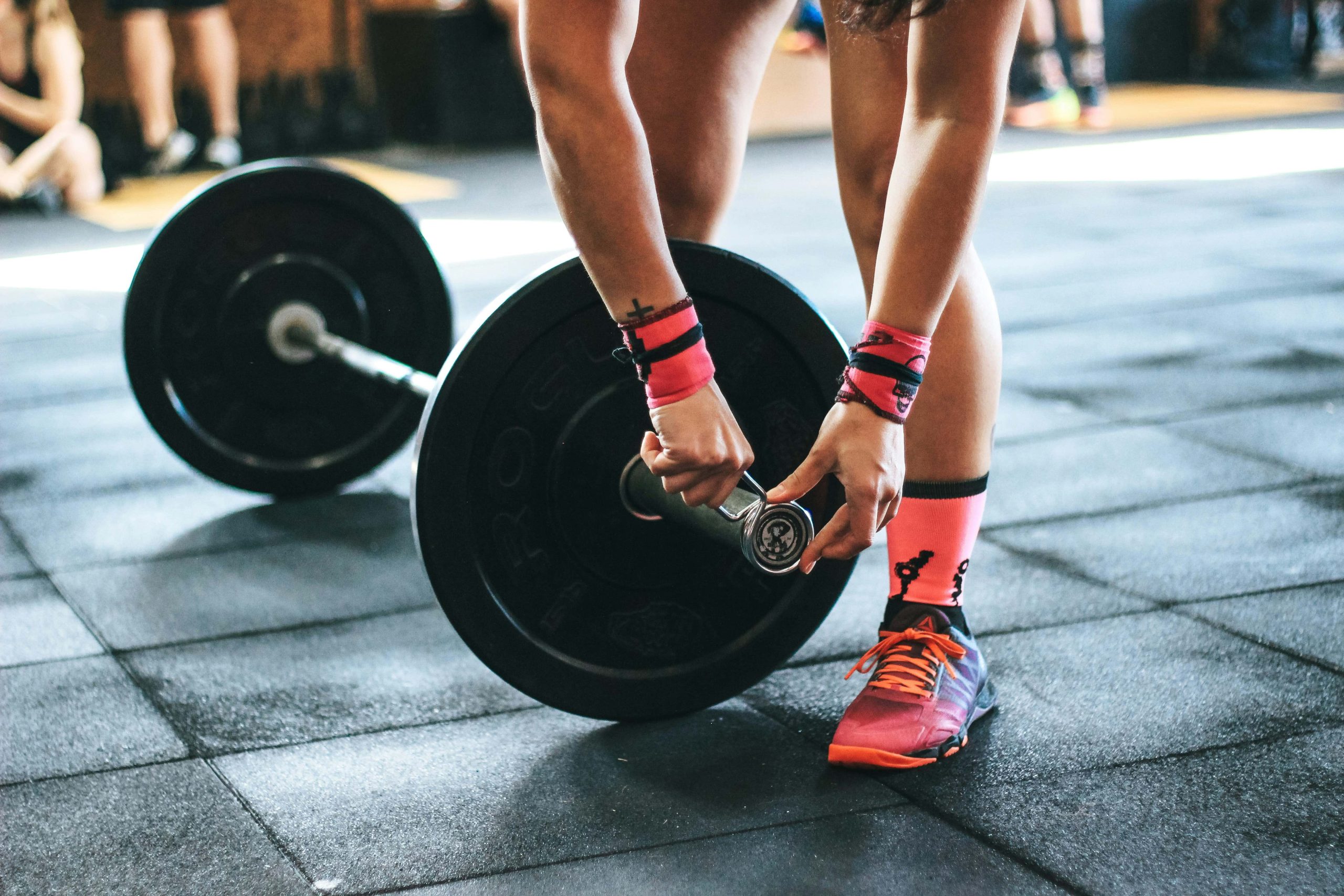
Between meetings, emails, and everything else on your plate, it’s easy to push workouts to the bottom of your to-do list. But if you want to stay energised, sharp, and healthy long-term, resistance training needs a spot on your calendar.
You’ve probably heard that your metabolism naturally slows down as you age — but here’s the truth: age isn’t the main problem. Research shows that most of the metabolic decline we blame on aging actually comes from losing muscle mass, not the passage of time.
Muscle is a high-maintenance tissue — it burns calories even while you’re sitting in a meeting or answering emails. But as we get older and become less active, we gradually lose that muscle, unless we intentionally work to keep it.
The good news? Resistance training helps you preserve and rebuild that lean muscle, keeping your metabolism strong, stable, and efficient — no matter your age.
We often think of metabolism as something we’re either blessed with or doomed by — that mysterious “engine” that controls how quickly we burn calories. But here’s the truth: your metabolism isn’t fixed. It’s shaped by your daily habits, and one of the most powerful tools you have to keep it running efficiently is resistance training.
Muscle = Metabolic Gold
Muscle is one of the most metabolically active tissues in your body. Even at rest, it burns more calories than fat does — and it plays a critical role in everything from blood sugar regulation to hormone balance.
Starting in our thirties, we naturally lose muscle mass (a process called sarcopenia), which can slow down metabolism and make it harder to maintain a healthy weight or energy level. Resistance training helps stop that decline and even reverses it by stimulating muscle protein synthesis — the process of building and repairing muscle tissue.
Your resting metabolic rate (RMR) is the number of calories your body burns at rest. More muscle = a higher RMR. That means even when you’re not working out, your body is burning more energy just to keep your systems running. Strength training doesn’t just burn calories while you’re doing it — it keeps working after you leave the gym.
Insulin Sensitivity & Metabolic Health
Resistance training improves insulin sensitivity, which means your body becomes better at using carbohydrates for fuel instead of storing them as fat. This reduces the risk of insulin resistance and metabolic disorders like type 2 diabetes. It also helps balance key hormones that impact metabolism, including cortisol and growth hormone.
It’s Not About Lifting Heavy Every Day
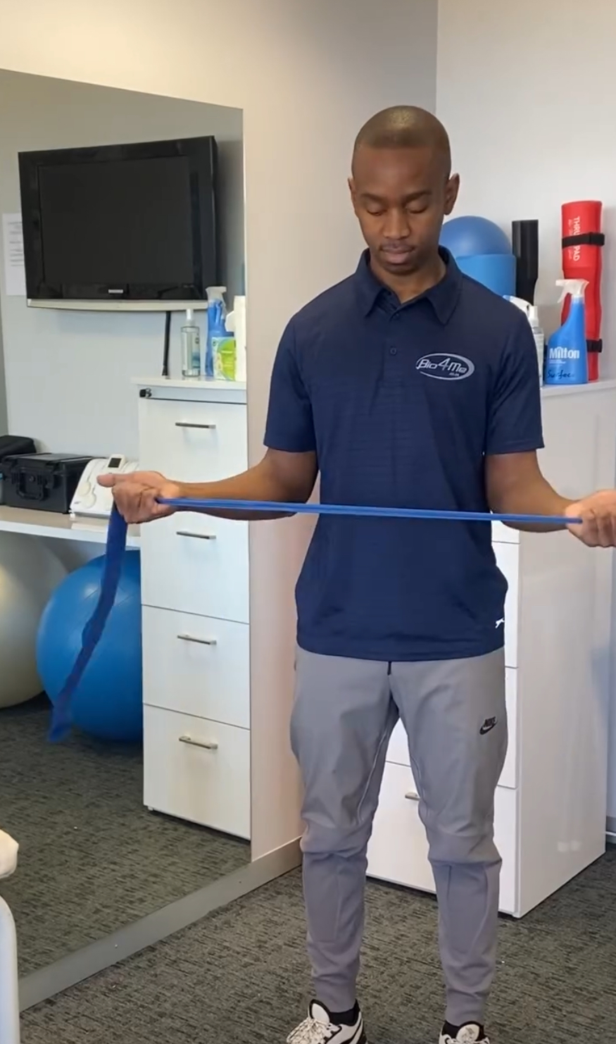
You don’t have to be a bodybuilder to reap the metabolic benefits. Bodyweight movements, resistance bands, dumbbells, or kettlebells all count. The key is progressive overload — gradually increasing the challenge over time to continue building and maintaining lean muscle.
Aim for 2–4 strength sessions per week, focused on full-body movement patterns (push, pull, squat, hinge, carry). Combine that with proper recovery and protein intake, and your metabolism will thank you.
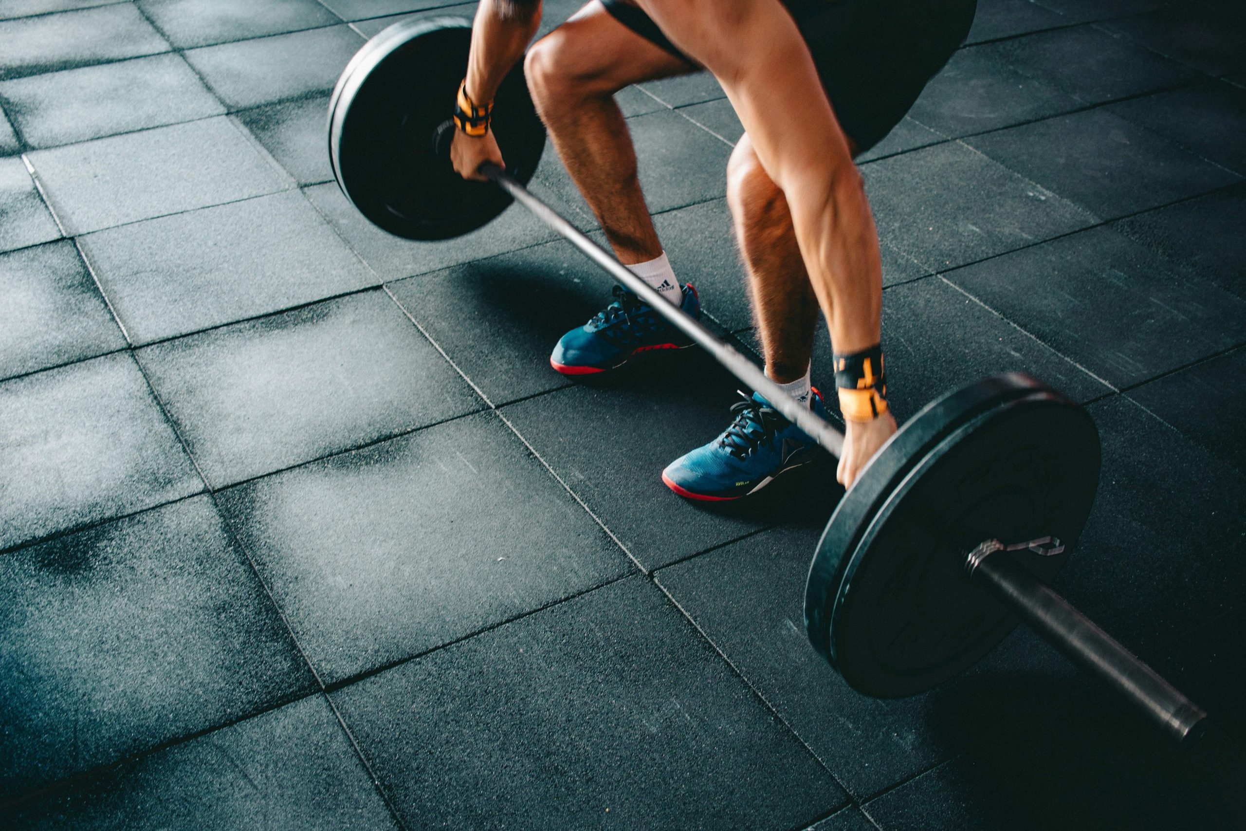
Prioritise compound movements — exercises that work multiple muscle groups. Examples: squats, lunges, rows, push-ups, presses, deadlifts.
Do 2–4 sets of 8–12 reps per exercise.
Rest 30–90 seconds between sets.
Cool Down (5 min): Stretch and breathe.
Focus on Progressive Overload:
To build and maintain muscle, your body needs to be challenged. Gradually increase the weight, reps, or difficulty over time. This is what keeps your muscles adapting and your metabolism humming.
No Gym? No Problem:
You can use resistance bands, dumbbells, kettlebells, or your own bodyweight (think push-ups, split squats, glute bridges). It’s the intensity and consistency that matter most — not fancy equipment.
Conclusion:
If you’re looking to maintain a strong, resilient body and a healthy metabolism as you age — resistance training isn’t optional. It’s essential.
So yes, cardio is great for heart health and mood. But if you want your metabolism to keep working for you, not against you? Pick up those weights.
If you require any advice on creating a personalised exercise program to improve your muscle mass and protect your metabolism, please do not hesitate to contact us!
Juvenile arthritis refers to a group of autoimmune and inflammatory conditions that cause persistent joint inflammation in children. The most common form is Juvenile Idiopathic Arthritis (JIA), which is diagnosed when arthritis lasts for at least six weeks and begins before the age of 16. Understanding juvenile arthritis is crucial for early diagnosis and effective management to ensure children maintain their quality of life.
The exact cause of Juvenile arthritis is completely understood but it is believed that genetics and environmental factors play a role in it. This condition is more common in girls.
Genetics: Family history of autoimmune diseases increases the risk.
Immune System: An overactive immune system mistakenly attacks the body’s own tissues.
Environmental Triggers: Infections or other external factors may trigger the onset in genetically susceptible individuals.
Symptoms
Joint Pain and Swelling: Persistent pain, swelling, and stiffness, especially in the morning or after periods of inactivity.
These focus on maintaining or improving joint flexibility.
Examples: Gentle stretching, yoga.
Strengthening Exercises:
Aim to build the muscles around joints to provide better support.
Examples: Resistance bands, light weightlifting, bodyweight exercises like squats.
Aerobic or Cardiovascular Exercises:
Improve overall fitness, heart health, and endurance.
Examples: Swimming (a great low-impact exercise), cycling, walking, dancing.
Balance and Coordination Exercises:
Help maintain balance and reduce the risk of falls or injuries.
Examples: Balance board exercises, standing on one leg, stability ball activities.
Water-based Exercises (Aquatic Therapy):
Particularly beneficial because the water’s buoyancy reduces the impact on joints while still providing resistance to build strength.
Examples: Swimming, water aerobics
Please feel free to contact us on social media or on our website if you need exercise rehabilitation and our friendly team would be more than happy to assist you.
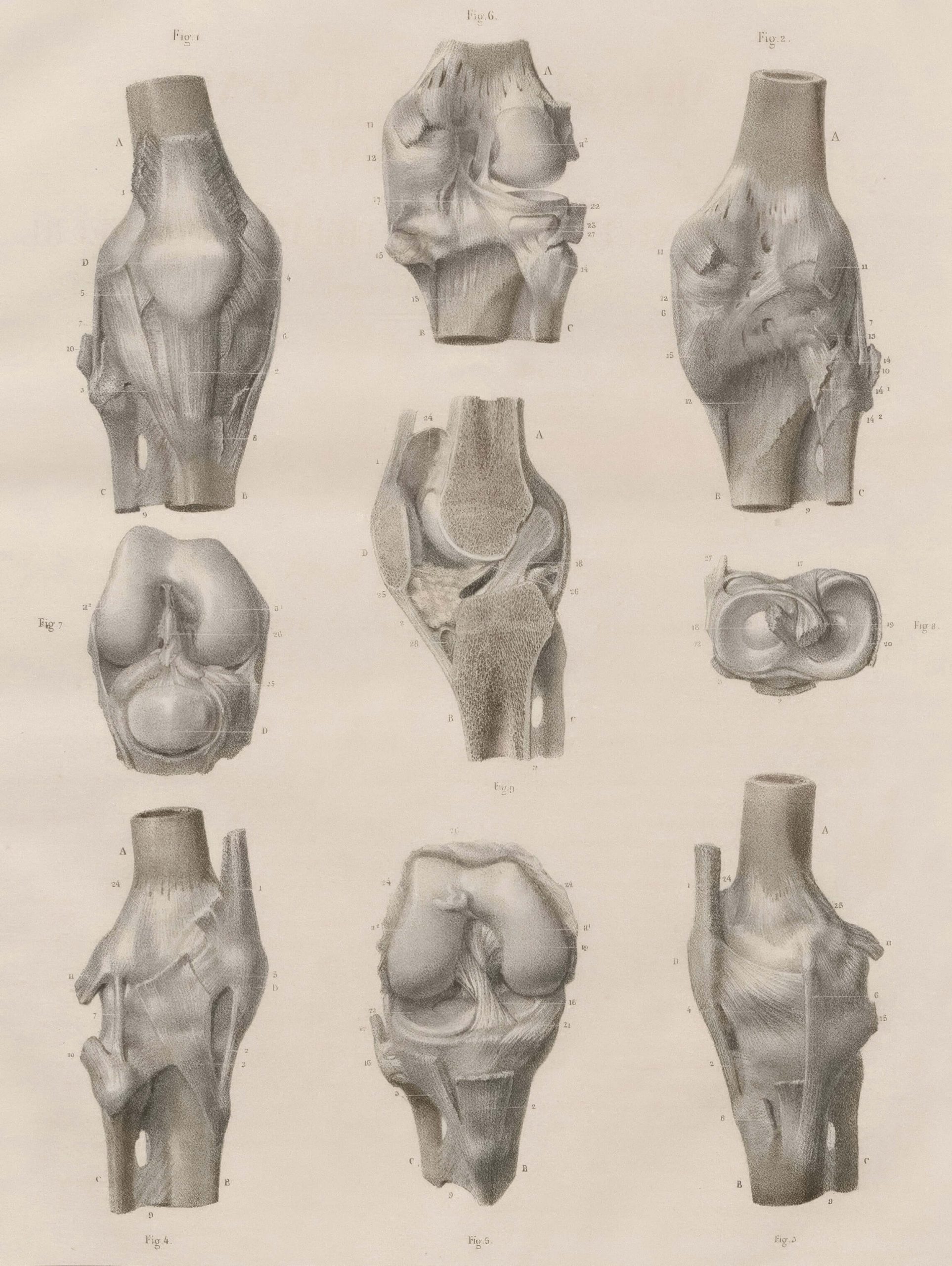
The posterior cruciate ligament is found in the knee just behind the anterior cruciate ligament (ACL). It connects the femur (thigh bone) to the tibia (shin bone) and it prevents the tibia from moving backwards. This ensures that there is smooth movement of the knee joint.
The PCL is comprised of robust connective tissues, including primarily collagen fibers, which are known for their high tensile strength. These fibers are crucial in anchoring the ligament firmly between the bones, ensuring optimal joint stability. The PCL’s primary role is to work together with other ligaments of the knee, such as the Anterior Cruciate Ligament (ACL), to stabilize the knee joint, particularly during dynamic movements such as running, jumping, and sudden directional changes.
PCL Injuries
Injuries to the PCL are often caused by direct trauma or excessive force to the knee. Common causes include motor vehicle accidents where the knee hits the dashboard, sports-related collisions, or falls that result in hyperextension of the knee. Risk factors for PCL injuries often include activities that involve high-impact forces or rapid changes in direction.
There are various types of PCL injuries, classified based on their severity. A Grade I injury involves a partial PCL tear, whereas a Grade II injury indicates a more significant but still partial tear. A Grade III injury is a complete tear of the PCL, and a Grade IV injury involves damage to the PCL as well as other knee ligaments
Symptoms of a PCL injury might include pain, swelling, a noticeable instability of the knee, and difficulty bearing weight. Diagnosis typically involves a combination of physical examination techniques, such as the posterior drawer test, and imaging studies like MRI to confirm the extent of the injury.
Treatment and Rehabilitation
Treatment for PCL injuries depends on the severity of the injury. Non-surgical approaches are often effective for less severe injuries (Grade I and II), and they include rest, ice, compression, and elevation (RICE) to reduce swelling and pain. Physical therapy exercises aimed at strengthening the quadriceps and hamstrings, which help stabilize the knee, are also critical. For more severe or complete tears (Grade III and IV), surgical intervention might be necessary. Surgical options include PCL reconstruction, where a graft is used to replace the damaged ligament. Rehabilitation plays an essential role in recovery post-surgery. This process typically involves a well-structured physical therapy regimen designed to restore knee function, improve strength, and regain full range of motion. The rehabilitation timeline can vary but often includes progressive exercises tailored to the individual’s specific needs and the injury’s extent.
Shoulder impingement syndrome (SIS) poses a significant challenge for athletes engaged in overhead sports. The intricate mechanics of the shoulder joint, combined with the repetitive stress of overhead movements, makes this condition prevalent among athletes. This Article delves into the different types of shoulder impingement, diagnostic approaches, and current management strategies to help athletes and healthcare professionals navigate this complex issue.
Types of Shoulder Impingement Syndrome
Subcoracoid ImpingementSubcoracoid impingement involves the compression of the anterior soft tissues of the shoulde. This is between the coracoid process and the lesser tuberosity of the humerus. This type of impingement can lead to significant discomfort and functional limitations, particularly in sports that involve repetitive forward motions.
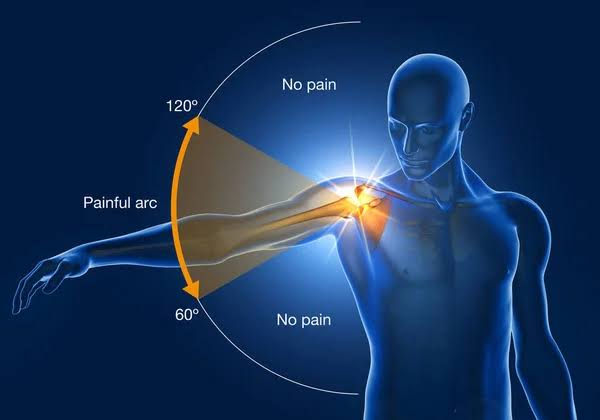
Subacromial Impingement Syndrome (SIS)Subacromial impingement occurs due to the extrinsic compression of the rotator cuff tendons. Generall, between the humeral head and the coracoacromial structures. It can also result from intrinsic degeneration of the supraspinatus tendon, which causes superior migration of the humeral head. This condition is frequently observed in athletes who engage in repetitive overhead activities, such as baseball pitchers or swimmers.
Internal ImpingementInternal impingement is a prevalent cause of shoulder pain in overhead athletes. It arises from the repetitive impingement of the articular surface. Generally during extreme abduction and external rotation of the arm. This type of impingement is particularly common among athletes performing high-intensity overhead motions.
Diagnostic Approach
Diagnosing shoulder impingement syndrome involves a thorough assessment that includes:
Sport-Specific Motion AnalysisUnderstanding the sport-specific movements that trigger symptoms is crucial. This helps in correlating the athlete’s pain with their specific athletic activities.
Physical Examination A combination of physical examination tests can enhance diagnostic accuracy. Tests such as the Neer test, Hawkins-Kennedy test, and the Jobe’s test can provide insights into the presence of impingement.
Radiographic Evaluation Radiographic imaging, including X-rays, ultrasound, or magnetic resonance imaging (MRI), is recommended to assess the extent of soft tissue abnormalities and structural damage. MRI is particularly useful in evaluating the rotator cuff tendons and the glenoid labrum.
Management Strategies
The management of shoulder impingement syndrome can be categorized into conservative and operative approaches, depending on the severity and chronicity of the symptoms:
Rest and Activity Modification: Reducing or modifying activities that exacerbate symptoms is essential.
Rehabilitation: A structured rehab program focusing on strengthening the rotator cuff muscles, improving scapular stability, and enhancing shoulder flexibility can be highly effective.
Medication: Nonsteroidal anti-inflammatory drugs (NSAIDs) or corticosteroid injections may be used to manage pain and inflammation.
Arthroscopic Surgery: In cases where conservative treatment fails, arthroscopic surgery may be considered. This may involve decompression of the subacromial space, repair of rotator cuff tears, or addressing any structural abnormalities contributing to impingement.
Conclusion
Shoulder impingement syndrome is a multifaceted condition that requires a comprehensive approach to diagnosis and management, particularly for athletes engaged in overhead sports. By understanding the different types of impingement, utilizing a thorough diagnostic approach, and applying appropriate management strategies, athletes can effectively address shoulder impingement and return to their sport with reduced pain and improved function.
Whether you’re a healthcare professional or an athlete
experiencing shoulder issues, feel free to contact us if you have any questions or concerns, or staying informed about the latest developments in the management of shoulder impingement syndrome is crucial for effective treatment and prevention of further complications.
Each year, more than 800 000 people are diagnosed with liver cancer all around the world. It is the leading cause of cancer-related deaths worldwide. Since 1980, the incidence of liver cancer diagnoses has tripled, whilst death rates have more than doubled. Exercise can play an important role in supporting individuals with liver cancer, both during treatment and in recovery. While exercise cannot cure liver cancer, it offers several significant benefits that can help improve the quality of life and overall health.
Liver cancer and its treatments (such as surgery, chemotherapy, or radiation) can cause fatigue, weakness, and muscle loss. Regular exercise helps maintain muscle mass, improve strength, and enhance overall physical function, making daily activities easier and reducing feelings of frailty.
2. Enhanced Immune Function
Moderate exercise has been shown to boost immune system function. A stronger immune system can help the body fight infections and potentially reduce the spread of cancer. This is especially important for liver cancer patients, whose immune systems might be weakened through cancer treatments.
3. Fatigue Management
Fatigue is one of the most common side effects of cancer and its associated treatments. Contrary to what one might think, regular, gentle physical activity can actually reduce cancer-related fatigue. Exercise increases energy levels and helps patients feel more alert and focused throughout the day.
4. Improved Mental and Emotional Well-Being
Being diagnosed with liver cancer can lead to stress, anxiety, and depression. Exercise releases endorphins, the body’s natural “feel-good” hormones, which can help reduce feelings of stress and improve mood. Physical activity can also provide a sense of accomplishment and control, boosting self-esteem and emotional resilience.
5. Better Circulation and Liver Function
Exercise promotes healthy blood circulation, which may help deliver oxygen and nutrients more efficiently to the body’s tissues, including the liver. This improved circulation supports liver function and overall body health, even in those with liver damage from cancer.
6. Maintenance of a Healthy Weight
Maintaining a healthy weight is important for liver cancer patients, as obesity and fatty liver disease are risk factors that can worsen liver health. Exercise, along with a healthy diet, can help control weight and reduce fat accumulation in the liver, potentially improving the body’s response to treatment.
7. Prevention of Treatment-Related Side Effects
Liver cancer treatments, especially chemotherapy, can cause side effects such as nausea, loss of appetite, and weakness. Regular exercise helps stimulate appetite, improves digestion, and reduces nausea, helping patients maintain their nutritional intake and avoid malnutrition during treatment.
8. Improved Cardiovascular Health
Cancer and its treatments can put additional strain on the cardiovascular system. Regular physical activity helps keep the heart and lungs strong, reduces the risk of blood clots, and supports cardiovascular health. This is especially important for patients undergoing treatments that may affect the heart, like certain chemotherapy drugs.
9. Support for Recovery and Rehabilitation
After surgery or during recovery, exercise can aid in rehabilitation by improving mobility, flexibility, and stamina. Gentle movement and stretching help prevent stiffness and promote healing, particularly after liver surgery or other invasive treatments.
10. Reduce Inflammation
Chronic inflammation can worsen cancer outcomes and affect overall health. Regular exercise has anti-inflammatory effects, helping to reduce systemic inflammation, which can positively impact liver health and potentially slow cancer progression.
11. Potential Improvement in Treatment Outcomes
Studies suggest that cancer patients who maintain regular physical activity during treatment may experience better outcomes, such as improved tolerance to chemotherapy or radiation and potentially longer survival rates. Exercise can enhance the body’s ability to tolerate treatments and recover more effectively.
Liver cancer patients should focus on gentle, low-impact exercises that support strength and stamina without causing undue stress. Some suitable forms of exercise include:
Walking: Low-impact and easy to incorporate into daily routines.
Swimming or water aerobics: Gentle on the joints and muscles, providing full-body conditioning.
Yoga or stretching: Improves flexibility, reduces stress, and promotes relaxation.
Strength training: Using light weights or resistance bands to maintain muscle mass.
Cycling: Low-impact cardio that supports cardiovascular health.
Precautions
Consult a doctor: Always consult with a healthcare provider before starting any exercise regimen, as liver cancer patients may have specific limitations due to treatment side effects or the condition of the liver.
Start slow: Begin with short, low-intensity sessions and gradually increase the duration and intensity based on your energy levels and how you feel.
Listen to your body: It’s important not to push too hard—rest when needed, and avoid exercise if you’re feeling particularly fatigued or unwell.
Conclusion
Exercise is a valuable tool in the holistic management of liver cancer, offering benefits that enhance physical, mental, and emotional well-being. When done safely and tailored to individual needs, physical activity can help liver cancer patients feel stronger, more energized, and better equipped to cope with the challenges of treatment and recovery.
If you require any assistance with regards to exercising to improve your liver cancer outcomes, please do not hesitate to contact us!
A groin hernia, also known as an inguinal hernia, occurs when a portion of tissue, such as part of the intestine, protrudes through a weak spot in the abdominal muscles in the groin area. This type of hernia is the most common, especially in men, though it can also occur in women.
Indirect Inguinal Hernia: This type is more common and occurs when the hernia sac enters the inguinal canal, often due to a congenital weakness. It is more common in males.
Direct Inguinal Hernia: This occurs when the hernia sac bulges directly through a weak spot in the abdominal wall muscles. It is usually related to aging and wear and tear.
Femoral Hernia: Though less common, this type occurs when tissue pushes through a weak spot near the femoral artery in the upper thigh/groin area. It is more common in women.
Causes:
Congenital weakness in the abdominal wall.
Strain.
Heavy lifting.
Chronic coughing or sneezing.
Obesity.
Pregnancy.
Treatment:
Watchful Waiting: Small hernias that aren’t causing symptoms might be monitored.
Surgery: The definitive treatment is surgical repair, which can be done via:
Open Hernia Repair (Herniorrhaphy): A traditional approach where the surgeon makes an incision in the groin to push the protruding tissue back in place and repairs the weakened area.
Laparoscopic Hernia Repair: A minimally invasive approach using small incisions and a camera to guide the repair.
Preventing a groin (inguinal) hernia involves strengthening the muscles in the abdominal and groin areas, improving core stability, and practicing safe lifting techniques. While exercise can help reduce the risk, it’s important to avoid activities that put excessive strain on these muscles. Here are some exercises that can help strengthen the muscles around the groin and abdomen:
1. Pelvic Tilts
2. Leg Slides
3. Wall Sits
4. Glute Bridges
Safe Lifting Techniques
When lifting heavy objects, bend at the knees, not the waist.
Keep the object close to your body.
Use your legs to lift, not your back.
These exercises can help strengthen the muscles and reduce the risk of developing a groin hernia, but it’s important to consult a healthcare professional before starting any new exercise routine, especially if you have any existing health concerns.
If you require any advice on creating a personalized exercise program to reduce your risk of suffering from a groin hernia, please do not hesitate to contact us!
Leukemia is a type of cancer that affects the tissues of the body that produce blood cells such as your bone marrow, lymph nodes and spleen. Leukemia mainly affects the white blood which are an important part of the immune system. The bone marrow will produce abnormal white blood cells that do not functional properly. Individual with leukaemia can benefit from exercise in their recovery journey.
Leukemia develops from genetic mutations in a single blood cell in the bone marrow, leading to the uncontrolled growth of abnormal white blood cells. These mutations can be triggered by factors like radiation, chemical exposure, or viral infections. As the mutated cell multiplies, it crowds out normal cells, disrupting blood production and immune function. This results in symptoms like anemia, infections, and bleeding disorders. In advanced stages, leukemia cells can spread to other organs and evade the immune system, leading to the progression of the disease, which can be acute and aggressive or chronic and slow developing.
The exact cause of Legg-Calvé-Perthes disease is not fully understood, but it is believed to involve a combination of genetic factors and vascular factors that affect the blood supply to the hip. Symptoms can include limping, pain in the hip or groin area, and limited range of motion in the hip joint. Over time, as the bone tissue heals and regrows and the body reabsorbs the dead bone tissue, the femoral head may become deformed and lead to arthritis in adulthood. This arthritis is caused by the remodelled femoral head being enlarged or flattened. It could take an individual 2 to 4 years to fully recover from this condition.
Treatment options vary based on the child’s age, the stage of the disease, and the degree of hip involvement. Here are common treatments for LCPD:
Rest and Activity Modification: Limiting weight-bearing activities to reduce stress on the hip joint.
Physical Therapy/Biokinetics: Exercises to maintain hip motion and strengthen the muscles around the hip.
Medications: Nonsteroidal anti-inflammatory drugs (NSAIDs) to relieve pain and reduce inflammation.
Bracing or Casting: Using braces or casts to keep the femoral head within the hip socket, promoting proper healing.
Surgical Interventions:
Osteotomy: Realigning the femur or pelvis to improve the fit of the femoral head in the hip socket.
Hip Distraction: Using an external fixator to allow the hip joint to heal by temporarily relieving pressure.
Observation: Regular follow-ups with imaging to monitor the progression of the disease.
Early diagnosis and treatment are crucial for better outcomes in LCPD. Consulting a paediatric orthopaedic specialist is essential for a tailored treatment plan.
Benefits of exercise Rehabilitation in LCPD
Maintains Joint Mobility:
Stretching and Range-of-Motion Exercises: These exercises help maintain and improve the range of motion in the hip joint, preventing stiffness and promoting better joint function.
Strengthens Muscles:
Strengthening Exercises: Strengthening the muscles around the hip can provide better support for the joint, reducing pain and improving mobility. Strong muscles also help distribute the weight more evenly across the hip joint.
Reduces Pain and Inflammation:
Low-Impact Activities: Activities like swimming or cycling can enhance blood circulation, reduce inflammation, and decrease pain without putting too much stress on the hip joint.
Prevents Muscle Atrophy:
Regular Exercise Routine: Ensuring that the muscles remain active prevents atrophy (muscle wasting), which is essential during the recovery phase of LCPD.
Improves Overall Physical Health:
General Fitness: Maintaining overall physical health through exercise helps in faster recovery and better management of the condition. It also promotes cardiovascular health, which can be beneficial for the overall healing process
A Biokineticist can design a personalised exercise program tailored to the individual’s specific condition and progression of LCPD. They can ensure exercises are done correctly and adjust the regimen as needed.
Blood pressure is measured in millimeters of mercury (mmHg) and recorded as two numbers: systolic pressure (the pressure when the heart beats) over diastolic pressure (the pressure when the heart rests between beats). Normal blood pressure is usually around 120/80 mmHg. Hypertension is defined as having a consistent reading of 130/80 mmHg or higher.
Aerobic Exercise: Activities like walking, jogging, cycling, and swimming are highly effective in lowering blood pressure. Aim for at least 150 minutes of moderate-intensity or 75 minutes of high-intensity aerobic exercise per week.
Resistance Training: Strength training exercises, such as lifting weights, can also help reduce blood pressure. Incorporate resistance training exercises at least two days a week.
Flexibility and Balance Exercises: While these exercises, such as yoga and stretching, do not directly lower blood pressure, they improve overall physical fitness and can reduce stress levels, which indirectly helps manage hypertension.
Weight Management: Physical activity aids in maintaining a healthy weight, reducing the strain on the heart and blood vessels.
Improved Blood Vessel Function: Exercise improves the elasticity of blood vessels, allowing them to expand and contract more easily, which helps lower blood pressure.
Stress Reduction: Regular physical activity reduces stress hormones, such as cortisol, which can contribute to high blood pressure.
Enhanced Kidney Function: Exercise helps kidneys better regulate sodium and fluid balance, contributing to lower blood pressure.
Getting Started
For those with hypertension, it’s essential to consult with a healthcare provider before starting a new exercise regimen. Thereafter, it is important to consult with a Biokineticist to guide you on the correct exercise and they will help you to gradually increase the intensity and duration of exercise. Consistency is key, and incorporating a variety of activities can keep the routine enjoyable and sustainable.
Conclusion
Exercise is a powerful tool in the fight against hypertension. Regular physical activity can help lower blood pressure, improve heart health, and enhance overall well-being. By making exercise a regular part of daily life, individuals can take significant steps toward managing their blood pressure and leading healthier, longer lives.