WITS Exercise Physiology
Posted on March 15th, 2012 by Andries Lodder

New WITS Exercise Physiology Lab study in Cyclists
by Aletta Esterhuyse
The Wits Exercise Lab has a new and very exciting study on comparing energy drinks ingested during cycling to investigate differences in energy metabolism and of cause cycling performance. This study will help you to understand what is truly the best mix for your racing fuel.
We invite male cyclists or triathletes who match all of the following characteristics to participate:
- Competitive male cyclists who are highly trained and include at least 12 hours cycling training per week.
- Cyclists who are generally healthy, not on medication and are non smokers
- Cyclists aged 18 to 45 years old
- Cyclists who are able to visit our laboratory for 5 separate 3 hour visits on a weekday arriving at 10 am (once a week).
The study’s focus is on comparing energy drinks ingested during cycling to investigate differences in energy metabolism and of cause cycling performance. In fact, in our quick releasing energy drink (or high glycaemic index mix) we hope to use very recent updated scientific recommendations that has not as yet been applied to off-the-shelve products. This study will help you to understand what is truly the best mix for your racing fuel.
We intend comparing the following 5 beverages:
- Placebo flavoured water
- High glycaemic index carbohydrate only (maltodextrin+fructose), i.e. your typical energy drink but with slightly more fructose
- Low glycaemic index carbohydrate (isomaltulose), i.e. a sugar used in some drinks such as 32GI and even USN Epic Pro
- High GI carbohydrate with added milk protein, casein hydrolysate, i.e. energy drink #2 with added protein (Peptopro)
- High GI carbohydrate with added milk protein, whey hydrolysate, i.e. energy drink #2 with different protein source, used in USN Epic Pro
We are looking for ten highly competitive male cyclists to take part. Each cyclist will first will undergo standard performance testing, to determine VO2max, lactate thresholds and peak power output. Each cyclist will then complete 5 separate experimental trial including a different energy drink during each trail. Each experimental trail will be separated by at least 1 week. During each trial you will cycle for 2 h at a fixed steady-state on our cycle ergometer for assessment of metabolism. This is immediately followed by a 16 km time trial on your own bicycle fitted to our Tacx ergometers that simulates a time trial route with real time video footage of the coarse. The programme we have chosen depicts you as an animated cyclist and looks similar to a play-station game only you have to pedal to get your character to move.
Please let us know if you are keen to be involved. We will start with performance testing mid-April and experimental trials soon after that.
You can either contact them directly or through Bio4Me and you will be provided with more details
Aletta: Aletta.esterhuyse@wits.ac.za
Tanja: oosthuyse@polka.co.za
Andries: send me an email
Office New Look
Posted on March 12th, 2012 by Andries
Importance of VO2max and Lactate Testing
Posted on February 6th, 2012 by Andries
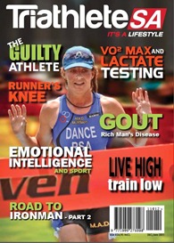
By Andries Lodder for TriathleteSA Magazine Dec|Jan 2012 Issue
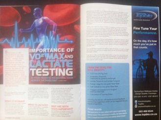
Is your training weakening your aerobic metabolism?
Blood pressure (BP) and cholesterol are used as predictors of potential disease, whereas fitness is a predictor of health. The healthier you are, the less chances you have of cardiovascular or respiratory diseases. We get our BP and cholesterol checked regularly, what should athletes be doing to check their fitness levels? VO2max and Lactate Tests accurately measure fitness and provide constructive feedback to athletes looking to increase their performance.
Why is this important for a runner, cyclist or triathlete?
Keep reading and you’ll soon find out.
What is VO2max?
VO2max is the maximum rate of oxygen consumed in millilitres one can use in one minute per kg of body weight. It is an important test for evaluating the cardiovascular capacity of an athlete and is the maximum capacity to transport and utilize oxygen during incremental exercise. It is also known as aerobic capacity, which reflects the physical fitness of a person. With an increase in oxygen consumption, you see cascades of positive events that all serve to increase the fuel economy of an athlete. Highly trained athletes generally have a higher VO2 max than untrained individuals.
What is Lactate?
Lactate Threshold testing is considered to be the single most important determinant of success in endurance related activities. Training at the right intensity is important to help prevent over or under training. During the demands of high intensity exercise, the cells utilise a substantial amount of glucose and glycogen (stored glucose). The by-product of the glucose breakdown is lactate. This increase in lactate coincides with an increase in blood and muscle acidosis, therefore lactate is an excellent indirect marker of muscular cellular fatigue or the “burn” sensation or that feeling of hitting the wall.
Why are both tests important?
Both VO2max and lactate turnpoint are valuable and independent physiological markers for your current state of fitness, in addition the gas analysis measurements will allow you to determine the point at which your body is using fat as the primary energy source as opposed to carbohydrates. The threshold calculations are crucial for development of accurate heart rate zones and training parameters. Utilising information from the VO2max and lactate tests can identify the most appropriate training intensity and type of training for you specifically rather than a handed down program from a mate or a predetermined heart rate zone program designed for the “general” population who are just interested in getting fit, but not focused on improving their best time or getting to the top of their game.
Athletes who make the common mistake of engaging in training structures that are much too intense are subject to reduced aerobic efficiencies. It is always a shocking discovery for any athlete to be confronted with the fact that their training has actually led to a systematic weakening of their aerobic metabolism. In other words, because they were unaware that the majority of their training was done in excess of their lactate threshold, there has been little stimuli for the body to increase its oxygen consuming abilities.
Because the data from a Vo2max and Lactate test will provide a detailed outline of whether fat or carbohydrates are being used most readily and to what extent, it is possible to determine, to the calorie, how many grams of carbohydrates are necessary to continue fuelling at a given intensity and avoid complete muscle glycogen deficit.
If you would like to improve your ability to deal with lactate or to work in a zone that will primarily use fat as an energy source, this test can provide the accurate parameters needed.
If you are a runner, you should be tested whilst running, if you are a cyclist, you should be tested on your own bike. If you are triathlete, you should be tested on both the run and cycle as the information between the two efforts will vary in some cases by quite a margin.
From the tests you will identify:
VO2 max (ml/kg/min)
- Anaerobic threshold (measured by gaseous exchange)
- Lactate threshold and lactate turnpoint
- Energy usage for any given heart rate
- Fuel mixture for any given heart rate
- Oxygen consumption at a range of exercise intensities
- Calculation of INDIVIDUALIZED training zones
- Following the test you will receive structured feedback and training suggestions which can be integrated into you regular programme.
Final words
The question is not what will I gain from doing these tests? The question is really by how much am I crippling myself by not doing these tests!
Ask an Expert: Backwards Running
Posted on February 6th, 2012 by Andries
By Andries Lodder for Modern Athlete Magazine Feb 2012 Issue
Backwards Running: Fact or Fiction?
Question:
I have heard so much about Backwards Running, but I’m not even sure what it is and if it is a fact or fiction.
Answer:
During locomotion the most involved joint of the lower body is the patello-femoral joint. Forming part of this joint is the patella, which is a sesamoid bone which reduces patello-femoral stresses, as well as increases the lever arm of the quadriceps muscles. The quadriceps muscle is the dynamic stabilizer of the patello-femoral joint. This joint is also the most common site for anterior knee pain, contributed by weakness of the quadriceps muscle.
Backwards running has numerous benefits which include burning one third more calories than forward running, it also develops better balance and stamina, it works the quadriceps muscles more than forward running, improves flexibility, reduces risk of injuries to the patello-femoral joint, you can still run while you are injured, improve leg speed and better performance, improved posture as well as your senses will be enhanced. It’s also stated that the volume of muscle active per unit of force applied to the ground was 10% greater when running backward than forward.
Normal forwards running contracts your muscles (quadriceps and hamstrings) mainly concentrically (muscles shorten during the contraction for force production phase of running) and eccentrically (muscles elongates for muscle recovery phase) at specific points in your stride. Backwards running works your muscles concentrically (recovery phase) and mainly eccentrically (force production phase) at opposite points of your strides. Eccentric contractions cause more muscle damage and needs a longer time period for recovery, but increases the muscle mass and strength. The benefit of training eccentrically is that when you go back to the conventional forwards running, your stability will be improved because of the muscles being trained in the opposite way and therefore your opposing muscle groups will provide some contraction while they are supposed to be relaxed, to provide more support to the contracting agonistic muscle.
A study at Stellenbosch University showed that the technique of backwards running actually improved your cardiovascular fitness. They used female students over a six-week period, training three times a week compared with a female group that stuck with their normal training regimes. The findings of the study were that the backward runners were found to have significantly decreased their oxygen consumption, therefore improving aerobically and lost 2.5% body fat.
Dangers of backwards running also exist. One of them is that when running backwards you can’t see anything in front of you or in the way of your path. Turning your head around will reduce the chances of falling or loosing your balance, but could lead towards running much slower as well as increase neck strains.
Considering the benefits and the biomechanics behind the patello-femoral joint, backwards running would reduce the occurrence of patello-femoral joint pain due to the strengthening of the quadriceps and the increased flexibility, which corresponds to the rehabilitation for treatment of anterior knee pain including hamstring stretching and VMO (vastus medialis oblique) strengthening.
Therefore my final verdict is that it is more fact than fiction.
To read this article on the Modern Athlete webpage, follow link: Ask an Expert: Backwards Running or buy the Feb issue today
Plantar Fasciitis
Posted on February 2nd, 2012 by Andries
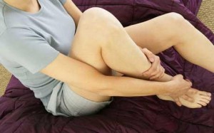
What’s that pain?
When your first few steps out of bed in the morning cause severe pain in the heel of your foot, you may have plantar fasciitis. It is one of the most common foot problems.
Plantar fasciitis is inflammation due to repeated overstretching of the plantar fascia ligament, usually at the point where the fascia is attached to the heel bone. This condition can also occur at the front of the foot. Tension develops in the plantar fascia both during extension of the toes and during depression of the longitudinal arch as the result of weight bearing.
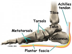
When a patient has plantar fasciitis, the plantar fascia becomes inflamed and worn out; these abnormalities in return can make normal activities quite painful. The pain typically worsens early in the morning after sleep. This pain is the result of the foot resting in plantar flexion (toes pointing forward) overnight. This allows the fascia to shorten. When you wake up in the morning the shortened or contracted fascia is stretched by simple movements and pain occurs and that is why you’re first few steps out of bed in the morning is painful. As you begin to loosen the plantar fascia, the pain usually decreases, but often returns with prolonged standing or walking
A Couple of signs or symptoms are pain in the anterior medial heel, usually at the attachment of the plantar fascia to the calcaneus (heel), as well as pain during the first few steps in the morning or after a long period of sitting.
The risk of developing plantar fasciitis are people who are active in sport, people that are flat-footed, middle-aged or older people, overweight individuals, incorrect footwear as well as pregnancy.
Some treatments used:
- Night splints. Use night splints to maintain a gentle, constant stretch across the plantar fascia.
- Heel pads. Felt, gel, or synthetic heel pads spread and absorb shock as the heel lands easing pressure on the plantar fascia.
- Decrease standing and stressful activities until heal pain is better.
- PRICE – pressure, rest, ice, compression and elevation would reduce inflammation.
- Stretch the heel cord and plantar fascia.
- Massage area of pain, especially in morning after a warm bath or shower.
For more details or questions on this topic, don’t hesitate to contact me on 0834686544 or send me an email
Contact Us
Please fill in your name, surname, email address and include a short message.
Osgood Schlatters Disease
Posted on January 26th, 2012 by Andries
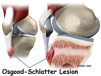
Overuse injuries are increasing among young athletes. This type of injury is the result of an increasing load on the musculoskeletal structures. The problem develops over time due to repeated stress and can be very painful and frustrating that leads to a chronic problem.
Osgood Schlatter disease: Inflammation and swelling at the site of insertion of the main quadriceps tendon at the top of the tibia (the tibial tubercle) just below the knee. It is common in
adolescence and result from excessive physical activity. Most cases resolve with time and rest.
It occurs most frequently in the younger athlete, due to the differences in structure and anatomy of the growing bone compared with that of the adult leg. The structural differences between the adult and growing bone is as follows:
- The articular cartilage of growing bone is thicker than in the adult bone, and the ability to remodel.
- The tendon attachments seat to the growing bone consists of relatively weak cartilage plate, the incidence of avulsion fractures may be increased.
- The metaphysis of long bones in children is more elastic and flexible, which causes the incomplete fractures (“greenstick” fractures) occurring only in children.
- With rapid growth phases extending the leg faster than the muscle and tendon can stretch. This causes temporary coordination and movement problems.
- Because of these differences, the younger athlete is more prone to cartilage and bone injuries, or a complete avulsion of the apophysis.
Osgood Schlatter presenting as anterior knee pain, usually associated with overuse or activity. The pain is felt by the attachment of the patella tendon to the tibial tubercle. The athlete will complain of pain during activities such as running, jumping, kneeling, squatting and up and down of stairs. In a sitting position with the knee in 90 ° flexion, a bony enlargement of the tibial tubercle is observed. Local tenderness over the anterior proximal tibial tubercle on palpation is a positive sign. The repeated irritation causes swelling, bleeding and gradual degeneration of the apophysis, which causes impaired circulation.
An effective examination of the lower limbs should be done to determine the extent of movement, particularly the flexibility of the hamstring and quadricep muscles. Muscle tests should be made to the strength in the knee and hip testing. The feet should also be tested for movement patterns that can lead to malalignment of knee injuries. Typical findings are external hip rotation, abduction movements and poor flexibility in the hamstring and quadricep muscles. Extension movements against a resistance will cause pain. Biomechanical tests that can be done include walking pattern, single and double leg squat (squat).
This condition is usually due to overuse. Overuse injuries are completely preventable for most young athletes. It is the responsibility of the parents and the coach to make sure that the young athlete’s program is not too demanding.Treatment is usually conservative. The goal of treatment is to reduce pain, increase strength and flexibility. Pain should be the main guideline to dictate the amount of activity.
Leg Length Inequality
Posted on January 25th, 2012 by Andries
By Dr. Bradley Waterer for Bio4Me
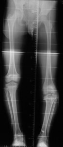
Is this leg longer?
Leg Length Inequality is extremely common. It may either be structural (where the bone in one leg is actually longer) or functional (where the problem is due to abnormal pelvic positioning).
Research indicates that Functional Leg Length Inequality (FLLI) exists in approximately 61% of the general population. Depending on the type and level of activity as much as 90% of an athletic team will have a Functional Leg Length Inequality. This condition predisposes people to acute and chronic recurring injuries involving ankles, knees, backs, necks and even shoulder and arm pain. One example of interest is that research found that 79% of people who experience lateral patella femoral pain have a Functional Leg Length Inequality. The condition almost always occurred in the “longer leg”.
If assessed correctly, Leg Length Inequalities can be identified to determine if a structural or functional abnormality is present. Once the problem is accurately identified the appropriate treatment you need can be administered.
For questions and enquiries contact Dr. Bradley Waterer (Chiropractor) on 0828029314 or visit http://www.sandtonchiropractic.com
Frozen Shoulder Rehabilitation Exercises
Posted on January 25th, 2012 by Andries
Wand Exercises

- Flexion: Stand upright and hold a stick in both hands, palms down. Stretch your arms by lifting them over your head, keeping your elbows straight. Hold for 5 seconds and return to the starting position. Repeat 10 times.
- Extension: Stand upright and hold a stick in both hands behind your back. Move the stick away from your back. Hold the end position for 5 seconds. Relax and return to the starting position. Repeat 10 times.
- External rotation: Lie on your back and hold a stick in both hands, palms up. Your upper arms should be resting on the floor, your elbows at your sides and bent 90°. Using your good arm, push your injured arm out away from your body while keeping the elbow of the injured arm at your side. Hold the stretch for 5 seconds. Repeat 10 times.
- Internal rotation: Stand upright holding a stick with both hands behind your back. Place the hand on your uninjured side behind your head grasping the stick, and the hand on your injured side behind your back at your waist. Move the stick up and down your back by bending your elbows. Hold the bent position for 5 seconds and then return to the starting position. Repeat 10 times.
- Shoulder abduction and adduction: Stand upright and hold a stick with both hands, palms down. Rest the stick against the front of your thighs. While keeping your elbows straight, use your good arm to push your injured arm out to the side and up as high as possible. Hold for 5 seconds. Repeat 10 times.
- Scapular range of motion: Stand and shrug your shoulders up and hold for 5 seconds. Then squeeze your shoulder blades back and together and hold 5 seconds. Next, pull your shoulder blades downward as if putting them in your back pocket. Relax. Repeat this sequence 10 times.
- Pectoralis stretch: Stand in a doorway or corner with both arms on the wall slightly above your head. Slowly lean forward until you feel a stretch in the front of your shoulders. Hold 15 to 30 seconds. Repeat 3 times.
- Biceps stretch: Stand facing a wall (about 6 inches away from the wall). Raise your arm out to your side and place the thumb side of your hand against the wall (palm down). Keep your elbow straight. Rotate your body in the opposite direction of the raised arm until you feel a stretch in your biceps. Hold 15 seconds, repeat 3 times.
Written by Tammy White, MS, PT, and Phyllis Clapis, PT, DHSc, OCS, for RelayHealth.
Published by RelayHealth.
Backwards Running Fact or Fiction
Posted on January 18th, 2012 by Andries
- Dixon, D. (2006). “The patello-femoral joint and impairment assessment.” Medico Legal. 26 July 2006. [Hyperlink http://www.medicolegal.com/newsletter]. 2 October 2007.
- Run the Planet (2000). “History of Backward Running.” Run the Planet. June 2000. [Hyperlink http://www.runtheplanet.com/resources/historical/backwards.asp]. 2 October 2007.
- Wright, S. & Weyand, P. G. (2001). The application of ground force explains the energetic cost of running backward and forward. The Journal of Experimental Biology, 204, 1805-1815.
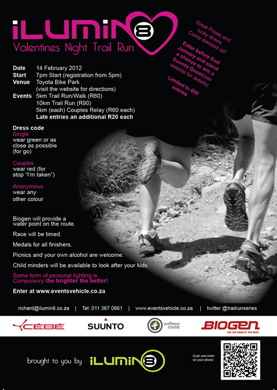
Valentines Night Trail Run
Posted on January 12th, 2012 by Andries

